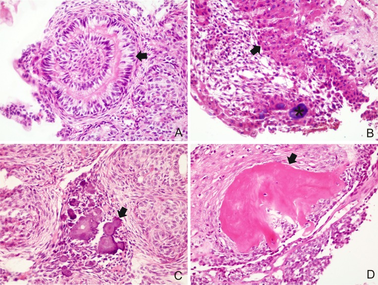
Fig. 3.
a Convoluted structures of tall columnar epithelial cells (arrow) (H&E ×400); b CEOT-like areas consisting of polyhedral, eosinophilic epithelial cells of squamous appearance with distinct cell boundaries and intercellular bridges (arrow) with calcified material (asterisk) (H&E ×400); c calcified material between the solid nests (arrow) (H&E ×400); d osteodentin juxtaposed to tumor cells (arrow) (H&E ×400)

