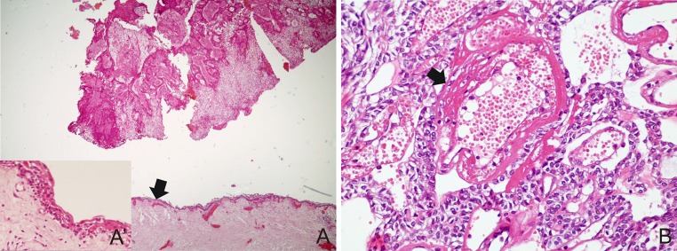
Fig. 4.
a AOT with area exhibiting thin non-keratinized stratified squamous lining epithelium mimicking a dentigerous cyst (arrow) (H&E ×40) and in detail (A′) (H&E ×400); b blood vessels of tumor stroma showing degenerative changes of endothelial lining and vessel wall (arrow), surrounded by cells with ovoid basophilic nuclei (H&E ×400)

