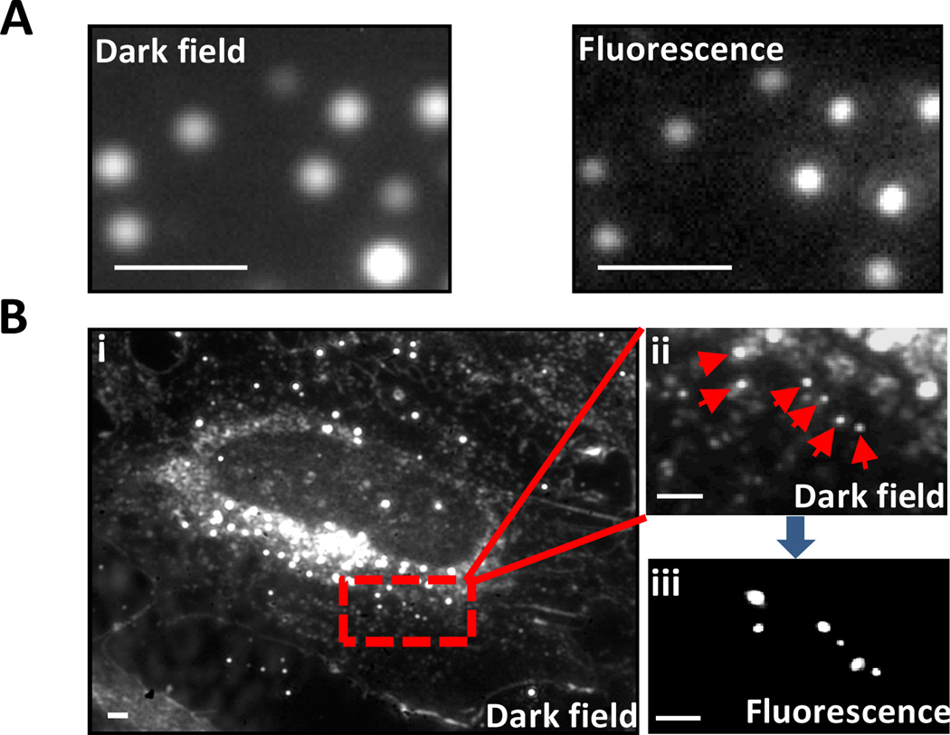
Fig. 3.
Dark field and fluorescence microscopy images of Au-NP-Pdots. (A) Dark-field (left) and fluorescence (right) images of Au-NP-Pdots immobilized on glass coverslip. (B) Dark-field and fluorescence images of Au-NP-Pdots inside cells. (i) Dark-field image of a whole cell; (ii) magnified image of the red rectangular area in (i); (iii) Corresponding fluorescence image of (ii). The Au-NP-Pdots were the same as those in figure 1A. Scale bar represents 1 µm.

