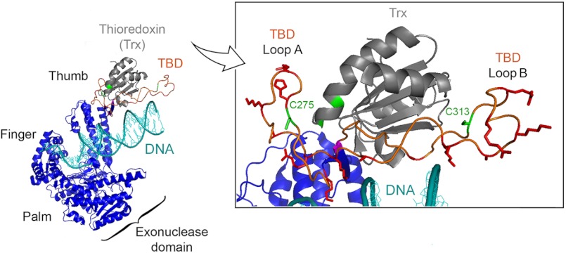
FIGURE 1.
Structure of T7 gp5/trx. The x-ray crystallographic structure of gp5/trx in complex with a primer-DNA template and a deoxynucleoside 5′-triphosphate was captured in a polymerization mode at 2.2 Å resolution (18). Primer-DNA template is shown in cyan. gp5 is present in blue with the three subdomains: palm, finger, and thumb. The three subdomains form a DNA binding groove leading to the active site containing the deoxynucleoside 5′-triphosphate. The processivity factor, trx depicted in gray, is bound to gp5 at a flexible loop of 76-amino acids, designated the TBD. The TBD, shown in orange, extends from the thumb. Inset, enlargement of the TBD and trx interface. In this structure, Cys-275 and Cys-313 on the TBD of gp5 and the active sites cysteines Cys-32 and Cys-35 of trx are highlighted in green. Thr-327 is highlighted in pink, whereas basic residues are highlighted in red.

