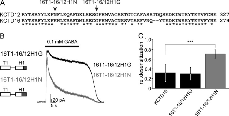
FIGURE 3.
Swapping experiments identify a region in the H1 domain that is critical for desensitization. A, sequence alignment of the C-terminal half of the H1 domains of KCTD12 and KCTD16. Identical and similar amino acids are marked with stars and dots, respectively. Arrows indicate the KCTD16/KCTD12 boundaries in the chimeric proteins 16T1–16/12H1N and 16T1–16/12H1G, which both lack the H2 domain of KCTD16. B, representative traces of GABAB-activated Kir3 currents recorded at −50 mV from CHO cells expressing GABAB(1b,2), Kir3 channels, and KCTD proteins. Kir3 currents exhibit strong desensitization in the presence of 16T1–16/12H1N but not 16T1–16/12H1G. C, bar graph summarizing the desensitization of Kir3 currents in the presence of KCTD proteins. Data are expressed as mean ± S.D.; ***, p < 0.001 compared with cells transfected with KCTD16 (Dunnett's multiple comparison test).

