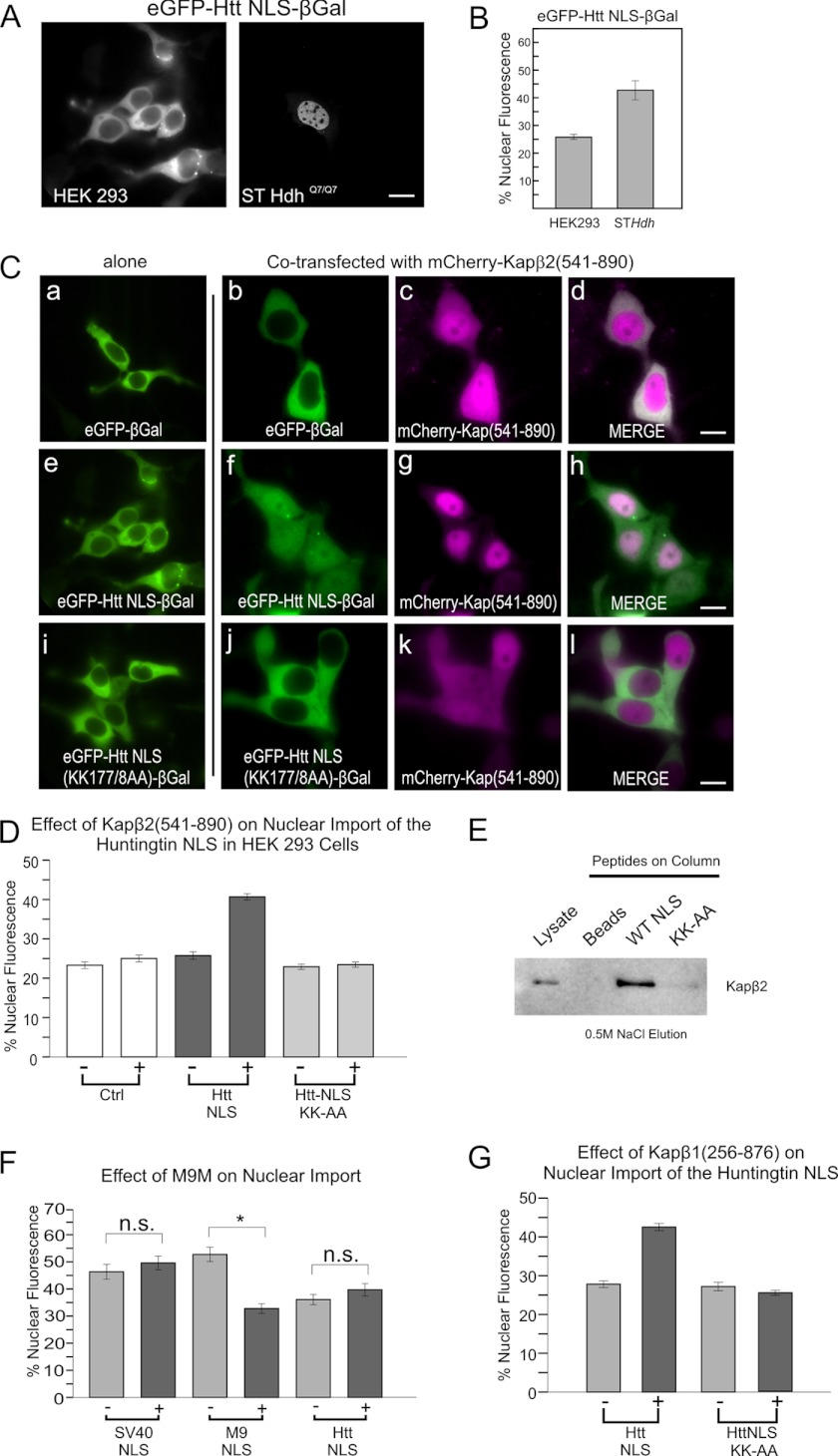
FIGURE 3.
Huntingtin PY-NLS utilizes the karyopherin β2/β1 import pathways. A, comparison of huntingtin NLS-mediated nuclear entry in human HEK293 versus mouse STHdh cells, quantified in B. C, overexpression of mCherry-karyopherin β2(541–890) (ΔRan binding) restoring wild-type NLS activity in HEK 293 cells (f–h), but not K177A/K178A mutant NLS (j–l). D, quantification of 3C. E, affinity chromatography of STHdh cell lysates on huntingtin NLS peptide-coupled columns. Column elutions at 0.5 m NaCl were separated by SDS-PAGE and Western blotted for karyopherin β2 (anti-Kapβ2,D45). Lysate lane contains 1/20 of column input. Beads indicates the beads-alone column. F, comparison of the SV40 NLS, M9 NLS, and huntingtin NLS in response to overexpression of M9M peptide competitor to karyopherin β2. G, overexpression of karyopherin β1(256–876) (ΔRan binding) increasing nuclear entry via the huntingtin NLS, but not a K177A/K178A mutant NLS. All NLSs are in GFP-β-galactosidase context in STHdh cells unless otherwise stated. For all quantification, n = ∼50 cells per construct. *, p < 0.01. Error bars, S.E.

