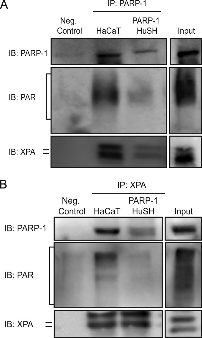
FIGURE 5.
Silencing PARP-1 protein leads to decreased association between PARP-1 and XPA. Co-immunoprecipitations were performed in HaCaT and PARP-1 HuSH cells. A, representative image of co-immunoprecipitations. PARP-1 was immunoprecipitated (IP) from cells, and the membranes were subsequently immunoblotted (IB) for PARP-1, PAR, and XPA. B, representative image of reciprocal co-immunoprecipitations. XPA was immunoprecipitated from cells, and the membranes were subsequently immunoblotted for PARP-1, PAR, and XPA.

