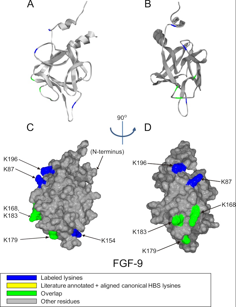
FIGURE 4.
Position of biotinylated peptides in FGF-9 (residues 52–208) identified by structural proteomics mapped onto the predicted three-dimensional structure (PDB 1IHK (42)). Labeled peptides are colored in blue, and overlapped peptides are colored in green. A and B, ribbon diagram. C and D, corresponding molecular surface. FGF-9 is shown using schematic representation. B and D, 90° left view of A and C.

