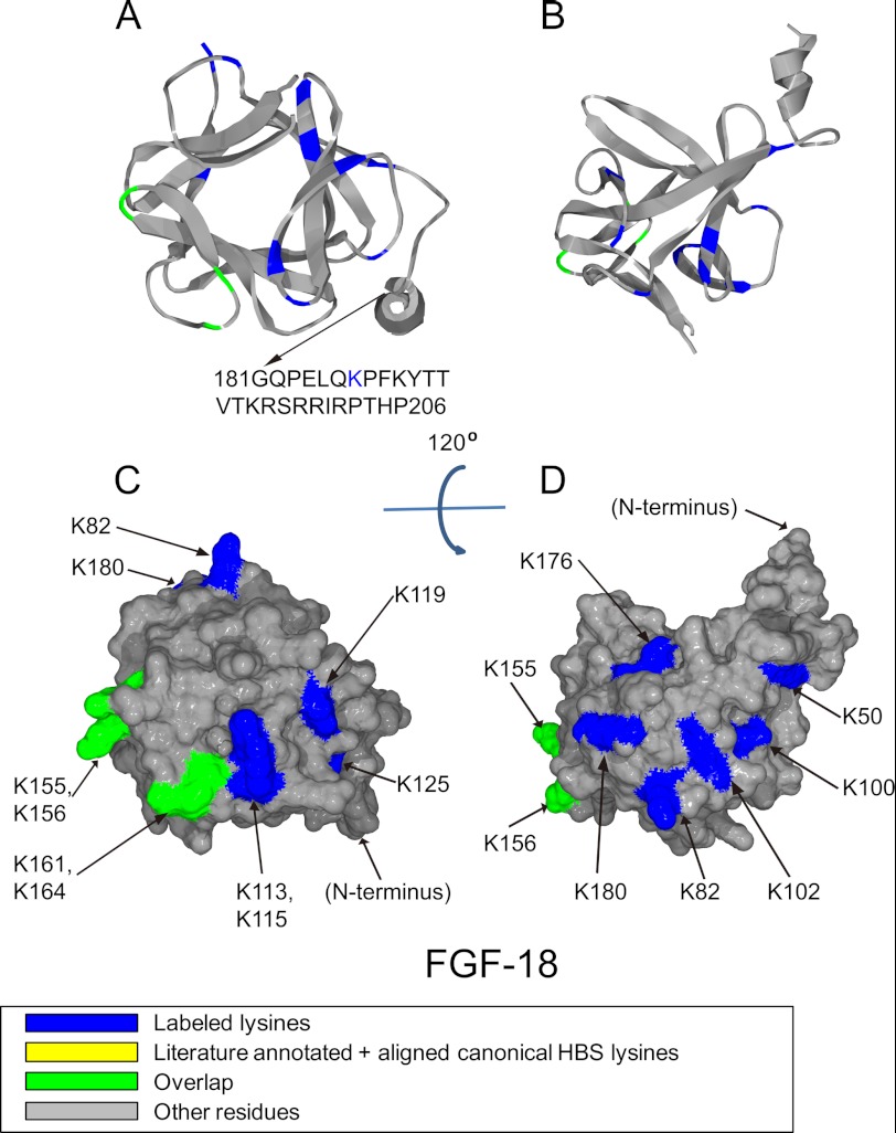
FIGURE 5.
Position of biotinylated peptides in FGF-18 (residues 35–176) identified by structural proteomics mapped onto the predicted three-dimensional structure (modeled structure from FGF-8b PDB 2FDB (56) using software SPDBV (57). Labeled peptides are colored in blue, and peptides overlapping with literature annotated and aligned canonical HBS lysines are colored in green. Loop between Gly-70 and Arg-71 was missing, and residues after Gly-181 was not shown due to model (FGF-8b) differences compared with FGF-18. A and B, ribbon diagram. C and D, corresponding molecular surface. FGF-18 is shown using schematic representation. B and D, 120° top view of A and C.

