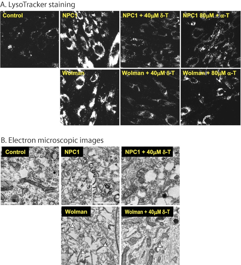
FIGURE 3.
δ-T and α-T reduced cellular acidic compartments and decreased pathological lysosomal inclusions in NPC1 and Wolman cells. A, the LysoTracker dye staining (for acidic compartments) was reduced in both NPC1 and Wolman cells by 40 μm δ-T or 80 μm α-T. The LysoTracker Red staining images were merged with Hoechst nuclei dye staining. B, electron micrographs of thin sections of NPC1 and Wolman fibroblasts were compared with control fibroblasts. The inset in the untreated NPC1 fibroblast shows in detail the lamellated and osmophilic structures in lysosomes (bar, 1 μm) that were significantly reduced by the treatment with 40 μm δ-T. In the untreated Wolman fibroblasts, the white arrowheads mark the typical elongated and cleft-shaped lipid droplets in lysosomes that were alleviated after the treatment with 40 μm δ-T.

