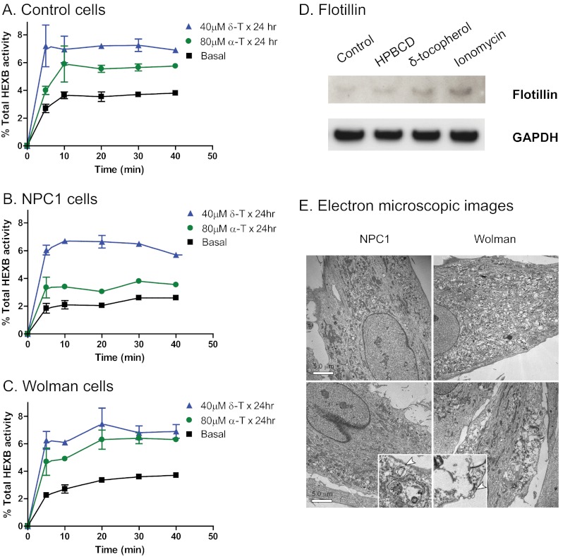
FIGURE 5.
Lysosomal exocytosis in δ-T- and α-T-treated NPC1 and Wolman disease fibroblasts. Lysosomal exocytosis was enhanced by δ-T and α-T in NPC1 (A) and Wolman (B) fibroblasts as well as in the control fibroblasts (C). After a 24-h pretreatment with 40 μm δ-T or 80 μm α-T, the activity of HEXB (a lysosomal enzyme) in extracellular medium significantly increased in all three cell types. The data shown are the mean of three experiments, and the error bars represent S.D. D, secretion of exosomes in δ-tocopherol-treated cells measured by Western blot analysis of flotillin 2 in the culture medium of control fibroblasts. The top panel exhibits the bands detected by an anti-flotillin 2 antibody in exosome fractions incubated with buffer, 2% 2-hydroxypropyl-β-cyclodextrin (a positive control), 160 μm δ-tocopherol, or 1 μm ionomycin. Equal loading was normalized by GAPDH levels in the cell lysate (bottom panel). E, electron micrographs of untreated NPC1 (top left) and Wolman (top right) cells versus treated NPC1 (bottom left) and Wolman (bottom right) cells show that the lysosomal storage materials in 40 μm δ-T not only decreased but also moved closer to the plasma membrane in overall distribution in the cell (bar, 5 μm). Insets, ×5 magnified areas with vesicles close to the plasma membrane.

