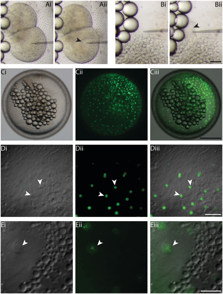
Fig. 2.
Microinjection technique and sytox green staining of YSN in K. marmoratus embryos. Using the agarose groove method, embryos can easily be microinjected in precise locations, as shown here with a two-cell (A [i and ii]) and late-blastula (B [i and ii]) injection. Despite the dark orange color of blastomeres, injected material can be tracked without the use of dyes (A [ii]; black arrowhead). To monitor fluorescence of YSN, sytox green (0.5 mM) was injected in the YSL at the late-blastula stage of development (B [ii]; black arrowhead). Fluorescence of YSN was examined in embryos still surrounded by their chorion, using a stereomicroscope (C [i–iii]), and at high magnification on an inverted compound microscope (×10 dry-objective lens) (D [i–iii]; white arrowheads). D (i) = DIC, D (ii) = fluorescence, D (iii) = overlay. To observe YSN at higher magnifications, the embryos were dechorionated and embedded in LMP agarose (×20 dry-objective lens) (E [i–iii]; white arrowheads). E (i) = DIC, E (ii) = fluorescence, E (iii) = overlay. Scale bars and associated images: 100 µm for A and B; 500 µm for C; 100 µm for D; and 50 µm for E.

