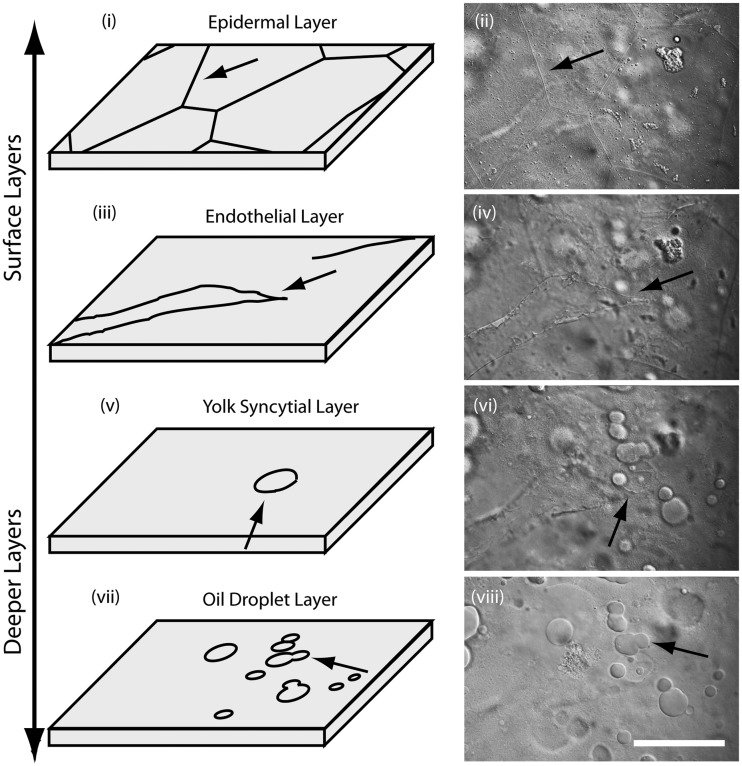
Fig. 3.
Imaging the surface layers of yolk in K. marmoratus embryos. During midsomitogenesis, geometrically shaped skin cells covered the surface of the yolk (i and ii; arrows). Under the epidermal layer, newly formed blood vessels were observed (iii and iv; arrows). The next deeper layer was the YSL where YSN were observed (v and vi; arrows). The deepest layer only contained oil droplets (vii and viii). Scale bar = 50 µm.

