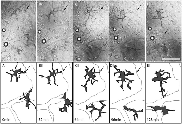
Fig. 4.
Time-lapse analysis of melanophores on the surface of the yolk in K. marmoratus embryos. (A [i])–(E [i]) DIC images of the endothelial layer on the yolk during the late embryonic development. (A [ii])–(E [ii]) Diagrams depicting positions and shapes of melanophores and vitelline vessels. A (i and ii) = 0 min; B (i and ii) = 32 min; C (i and ii) = 64 min; D (i and ii) = 96 min; and E (i and ii) = 128 min. Extending protrusions from the melanophores appear to come in contact with the edge of a vitelline vessel (A [i and ii]; B [i and ii]; C [i and ii]; arrows) and then detach (D [i and ii]; arrows) to touch another edge of the vitelline vessel (E [i and ii]; arrows). Scale bar = 50 µm.

