
Fig. 1.
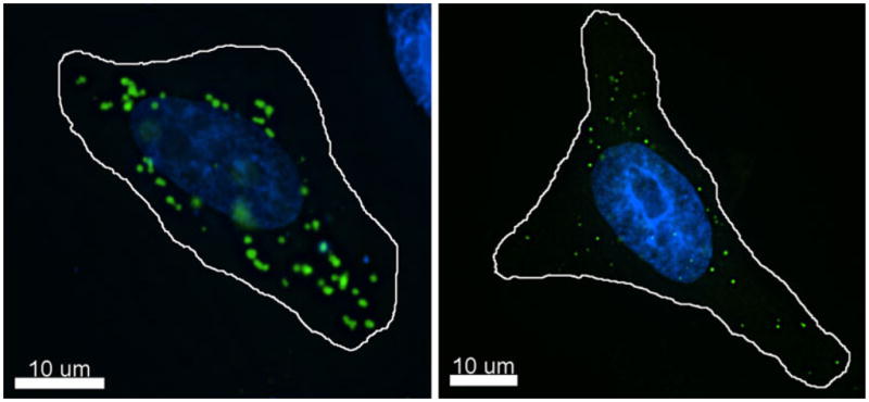
Subcellular localization of rhesus macaque TRIM5α. A, HeLa cells were seeded on glass coverslips and transiently transfected with YFP–rhTRIM5α using a PEI protocol. Z–stack images were collected and de-convolved using Deltavision deconvolution software. Individual channel images were superimposed to create the merged image. B, HeLa cells stably expressing YFP–rhTRIM5α from an integrated retroviral vector were seeded on coverslips. Z–stack images were collected and de-convolved using Deltavision deconvolution software. Individual channel images were superimposed to create the merged image