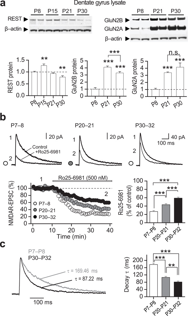
Figure 2. Transient increase in REST precedes the switch in NMDAR phenotype in rat dentate gyrus.
a, Representative immunoblots (top) from microdissected dentate gyrus tissue at indicated postnatal (P) ages, showing the developmental expression for REST, GluN2A, GluN2B and β-actin (see full-length blots in Supplementary Fig. 12). All values were normalized relative to P8. Quantification (bottom) showing that, as in the hippocampal formation (see Fig. 1), REST expression in dentate gyrus is transiently increased at P15 (bottom left panel), the NMDAR subunits GluN2B declines (bottom middle panel) and GluN2A increases (bottom right panel) (n = 4). b, Averaged sample traces of NMDAR-EPSCs (top) before and after bath application of the selective GluN2B antagonist Ro25–6981 (500 nM) at indicated postnatal ages (P7–8, P20–21, P30–32). Time course and summary data (bottom) showing that sensitivity to Ro25–6981 decreases progressively with age (P7–8, 2 animals/7 cells; P20–21, 2 animals/8 cells; and P30–32, 2 animals/6 cells). Averaged sample traces were taken at times indicated by numbers on the summary plot. c, Normalized NMDAR-EPSCs traces at different postnatal ages and summary data showing that the decay kinetics of NMDARs increases progressively with age. Summary data represent the mean ± s.e.m. **p<0.01; ***p<0.001; n.s., not significant.

