
Fig. 2.
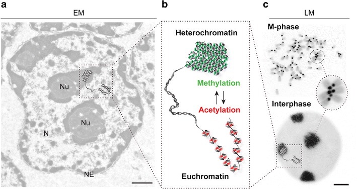
Heterochromatin: in need of definition? Historically and from a cytological point of view, Emil Heitz (see Fig. 1) distinguished hetero and euchromatin. a Within an exemplary electron microscopy (EM) picture (left) of a mouse liver cell nucleus (N nucleus, Nu nucleolus, NE nuclear envelope), heterochromatin appears as electron dense in contrast to the more open state of euchromatin. b With the recent advent of high-throughput epigenomics, molecular features (histone and DNA modifications) have been assigned to particular chromatin states and are shown in the simplified graphic enlarged in the center. c The cell cycle dynamics and cytological organization of the very condensed chromatin structures around the centromeres can be appreciated in the fluorescence light microscopy (LM) pictures (right) of M phase and interphase cells. FISH-stained mouse metaphase chromosomes and interphase cell with probes against pericentric heterochromatin (black) and DNA counterstaining (gray) are shown. Condensed pericentric heterochromatin regions from multiple chromosomes cluster together in the interphase cell nucleus forming the so-called “chromocenters.” Cytological and molecular definitions have not yet been conclusively linked together. Scale bars EM 0.5 μm and LM 2 μm