
Fig. 2.
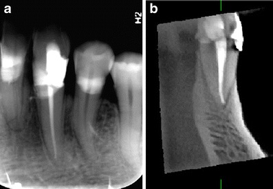
a A radiograph shows a short root filling in tooth 33 that is 2.5 mm short of the radiographic apex. b A sagittal CBCT view. Note the flush filling
Official websites use .gov
A
.gov website belongs to an official
government organization in the United States.
Secure .gov websites use HTTPS
A lock (
) or https:// means you've safely
connected to the .gov website. Share sensitive
information only on official, secure websites.
a A radiograph shows a short root filling in tooth 33 that is 2.5 mm short of the radiographic apex. b A sagittal CBCT view. Note the flush filling