Fig. 3.

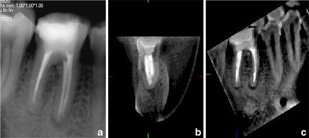
a A periapical radiograph of tooth 46. No void was observed in the distal root filling, but in the coronal and sagittal CBCT views (b, c), void was diagnosed

a A periapical radiograph of tooth 46. No void was observed in the distal root filling, but in the coronal and sagittal CBCT views (b, c), void was diagnosed