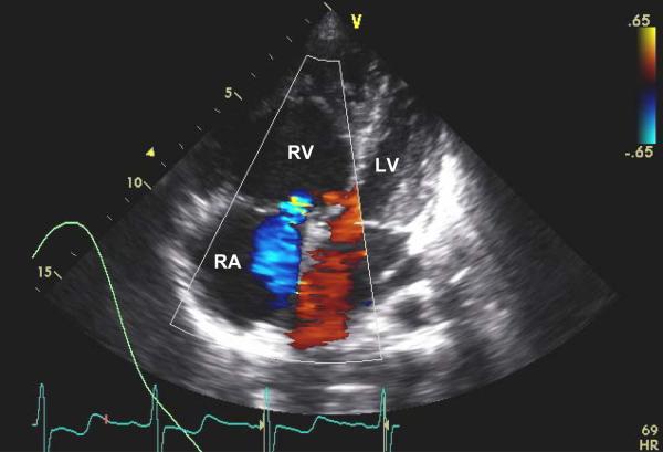
Image 2.
Two dimensional color flow Doppler image showing a subcostal four chamber view during systole from the transthoracic echocardiogram. The color flow Doppler shows severe tricuspid regurgitation (with the transducer present at the top of the image, the blue area in the right atrium indicates blood flow from the right ventricle back into the right atrium). The image also shows severe dilation of the right atrium and right ventricle with flattening and bowing of the intraventricular septum into the left ventricle. Normally, the right atrium and right ventricle should be smaller than the corresponding structures on the left during systole. RA = right atrium; RV = right ventricle; LV = left atrium; and V = position of the echocardiographer's transducer.

