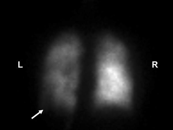
Image 5.
PA view of the nuclear medicine perfusion scan of the lungs after intravenous injection with the technetium 99m MAA isotope. The perfusion scan activity is significantly less on the left compared to the right. This image also shows a major perfusion defect in the lateral basal segment of the left lower lobe as indicated by the arrow. Also observed are smaller partial perfusion segment defects in the left upper lobe, the lingula, and the right upper lobe. The pulmonary ventilation perfusion scan was reported to have an intermediate to high probability for the presence of pulmonary emboli. L = left and R = right.

