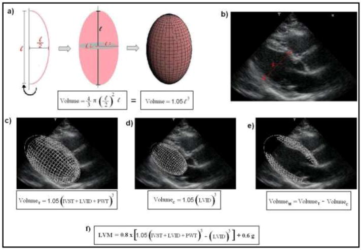
Figure 1. Principles for the assessment of LVM by echocardiography, as recommended by the American Society of Cardiology (ASE).20 Images from ‘c)’ to ‘e)’ refer to a schematic representation of the steps for the estimation of left ventricular myocardial volume, as initially proposed by Devereux et al.28 LVM is then calculated multiplying the myocardial volume by the specific gravity of myocardium (1.05 g).
a) A prolate ellipsoid of revolution, or prolate spheroid, is a three-dimensional figure formed by revolving an ellipse about its major axes. The ASE-recommended formula assumes that the left ventricle has a prolate ellipsoid of revolution shape, with minor radii that are half the major radius.
b) Schematic representation of the linear measurements for the assessment of LVM by echocardiography (para-esternal view), according to the ASE recommendations. 1-interventricular septum thickness (IVST); 2- Left ventricular internal dimension (LVID); 3- posterior wall thickness (PWT).
c) Calculation for the total left ventricular volume (VolumeT).
d) Calculation for the left ventricular internal cavity volume (VolumeC).
e) Calculation for the left ventricular myocardial volume (VolumeM).
f) The current ASE-recommended formula for the assessment of LVM. It is based in the initial concepts, but includes correction factors derived from regression analysis.20,35

