
Figure 2. Images from cardiac magnetic resonance (CMR) of two patients with Chagas cardiomyopathy. Case 1 has preserved cardiac geometry, but case 2 shows left ventricular remodeling. The usual assessment of LVM (LVM) by CMR does not require cardiac geometry assumptions, in opposition to linear measurements used in echocardiography. (Courtesy of Dr. Gustavo Volpe).
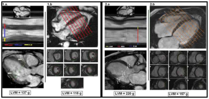
For 1.a and 2.a - CMR-derived images representing usual echocardiography views for linear measurements assessing LVM. ASW corresponds to interventricular septal thickness; EDD corresponds to left ventricular internal dimension; and PLW corresponds to posterior wall thickness. At the bottom, the ASE-recommended formula was used to calculate LVM (see figure 1 for full description).
For 1.b and 2.b - Usual CMR assessment for LVM, using contiguous short-axis slices covering the entire left ventricle from the atrio-ventricular ring to the apex (1 to 9). The estimated LVM is displayed at the bottom.