Figure 5.
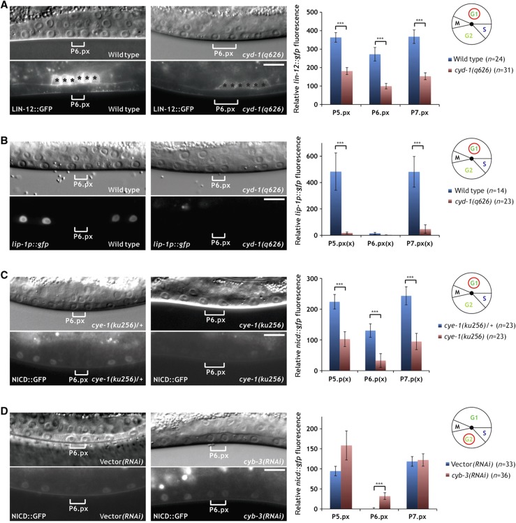
Differential regulation of NOTCH signaling by G1 and G2 cyclins. (A) Localization of LIN-12::GFP to the apical plasma membrane is diminished in the VPC descendants in cyd-1(q626) mutants (right panels) when compared with wild-type controls (left panels). Black asterisks denote uterine LIN-12::GFP expression. (B) Expression of the 2° fate marker lip-1p::gfp is reduced in the P5.p and P7.p descendants in cyd-1(q626) mutants (right panels) compared with wild-type controls (left panels). (C) NICD::GFP expression is reduced in the VPC descendants in homozygous cye-1(ku256) mutants (right panels) when compared with heterozygous cye-1(ku256)/+ controls (left panels). (D) NICD::GFP expression persists in the P6.p descendants of a cyb-3 RNAi-treated animal at the Pn.px stage (brackets in the right panels). Note that in contrast to cdk-1 (Figure 4C), cyb-3 RNAi did not induce a cell-cycle arrest. An empty vector RNAi-treated control animal is shown in the left panels. In all panels, the corresponding Nomarski images are shown on top. The scale bars represent 10 μm. Quantifications of LIN-12::GFP, lip-1p::gfp and NICD::GFP expression are shown to the right. Error bars indicate the standard error and asterisks indicate the significance as described in Materials and methods. Source data is available for this figure in the Supplementary Information

