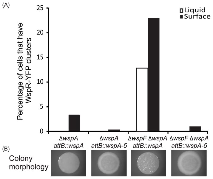
Fig. 9. Functional analysis of WspA and WspA-5.
(A) Ability of WspA/WspA-5 to phosphorylate WspR, measured by percentage of cells in the population that have at least one WspR-YFP cluster. Liquid grown cells: white bars; surface grown cells: black bars. (B) Colony morphology of corresponding strains. Vector control strains were ΔwspA attB::vector control and ΔwspFΔwspA attB::vector control, these strains did not have any WspR-YFP clusters and had smooth colony morphology (data not shown).

