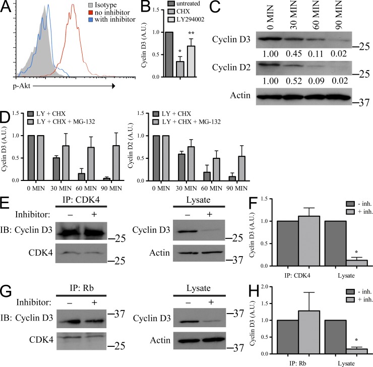
Figure 2.
Soluble cyclin D3, but not that associated with CDK4 and Rb, is regulated by PI3K. (A) Rag2−/− pro–B cells were cultured in IL-7 ± LY294002 for 60 min, fixed, permeabilized, stained for intracellular p-Akt, and analyzed by flow cytometry. (B) Rag2−/− pro–B cells were cultured in IL-7 alone (untreated) or with CHX or LY294002 for 60 min. Samples were prepared in NP-40 lysis buffer, resolved by SDS-PAGE, and membranes were probed for cyclin D3 and Actin. Expression of cyclin D3 was normalized to Actin, standardized as a ratio of the treated sample relative to the expression level in untreated samples (arbitrary units, A.U.), and reported here graphically as the mean ± SD, n = 3. Compared with the untreated sample: *, P < 0.001; **, P < 0.05. (C) Rag2−/− pro–B cells were cultured in IL-7 with LY294002 and CHX for the times indicated. TCLs were prepared and resolved as in B and membranes were probed for cyclin D3, cyclin D2, and Actin (representative experiment, n = 3). Relative molecular mass (kD) is as indicated. Cyclin expression reported below immunoblot in arbitrary units (A.U.) as described in B. (D) Rag2−/− pro–B cells were cultured in IL-7 with LY294002 (LY) and CHX or LY, CHX, and proteasome inhibitor MG-132 for the times indicated, analyzed as in C, and reported here graphically as mean ± SD, n = 3. (E–H) Rag2−/− pro–B cells were cultured in IL-7 with or without inhibitors LY294002 and CHX for 60 min. (E and G) CDK4 (E) or Rb (G) immunoprecipitates and corresponding TCLs were resolved by SDS-PAGE and membranes probed with indicated antibodies. IP lanes: 8 × 10⁶ cells; lysate lanes: 7 × 10⁵ cells. Relative molecular mass (kD) is as indicated. (F and H) Quantification of cyclin D3 from blots normalized to CDK4 (IP lanes) or Actin (TCL lanes), standardized as a ratio of the treated sample relative to the expression level in untreated samples (arbitrary units, A.U.) and shown as mean ± SD, n = 3. *, P < 0.05.

