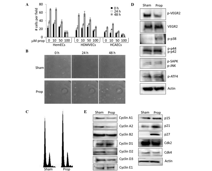
Figure 2.
Propranolol decreases the proliferation of human infantile hemangioma endothelial cells (HemECs). (A) HemECs, human dermal microvascular endothelial cells (HDMVECs), and human coronary artery endothelial cells (HCAECs) were treated with a dose curve of propranolol (0 to 100 μM) and cell proliferation was measured by counting changes in the number of cells/defined vision field over a 48-h period. (B) Time lapse microscopy image of sham and 50 μM propranolol treated HemECs over a 48-h period. (C) DNA content analysis of propidium iodide stained HemECs treated with sham or 50 μM propranolol for 48 h. (D) Western blot analysis detecting the levels of phosphorylated and total vascular endothelial growth factor receptor-2 (p-VEGFR-2 and VEGFR-2, respectively) and the phosphorylated forms of p38 (p-p38), p44 (p-p44), p42 (p-p42), stress activated protein kinase (p-SAPK), c-jun N-terminal kinase (p-JNK), and activating transcription factor 4 (p-ATF4) in HemECs treated for 24 h with sham or 50 μM propranolol. Actin levels were used as a loading control. (E) Western blot analysis detecting the levels of cyclins, cyclin dependent kinases, and cyclin dependent kinase inhibitors in HemECs treated for 24 h with sham or 50 μM propranolol. Actin levels were used as a loading control. Prop, propranolol.

