Figure 4.
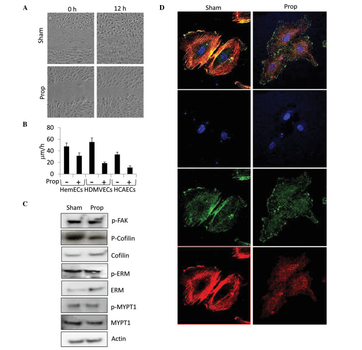
Propranolol disrupts HIHEC migration and actin cytoskeleton dynamics. (A) Confluent monolayers of human infantile hemangioma endothelial cells (HemECs) were scratch wounded and treated with sham or 50 μM propranolol. Progress of migration was monitored using time lapse photography over a period of 12 h. (B) Quantification of the speed (μm/h) of HemECs, human dermal microvascular endothelial cells (HDMVECs), and human coronary artery endothelial cells (HCAECs) treated with sham or propranolol from the time lapse images of the scratch assay. (C) Western blot analysis detecting the levels of the total and phophorylated (p-) forms of focal adhesion kinase (FAK), cofilin, ezrin/radixin/moesin (ERM), and myosin phosphatase-targeting subunit 1 (MYPT1) in HemECs treated with sham or 50 μM propranolol for 48 h. Actin levels were used as a loading control. (D) Confocal immunofluorescent imaging of sham or 50 μM propranolol-treated HemECs co-stained with Rhodamine conjugated phalloidin (red), DAPI (blue), and antibodies against phospho-FAK. Prop, propranolol.

