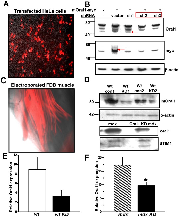
Figure 2. Effective knockdown of endogenous Orai1 gene in both wt and mdx FDB fibers by shRNA probe.
(A) Superimposed transmission light and red fluorescent images (100x) of Hela cells after 24 h transfection with shOrai1 with a RFP marker and mouse full length Orai1 with a myc tag (mOrai1-myc). (B) Western blot showing effective knockdown of the exogenous mOrai1 gene by shOrai1 probes (sh1∼3) No. 2 and No. 3. β-actin was used as a loading control. (C) Fluorescence image (100x) showing the successful transfection of FDB muscle 2 weeks after electroporation of shOrai1 probe. (D) Single fiber western blots confirmed the effectiveness of shOrai1 in knocking down the endogenous mOrai1 in wt mice (upper panel) and mdx mice (lower panel). Pooled extracts from 15 individual FDB fibers were loaded per lane. The exposure used to generate these images was adjusted to produce a robust signal that could be used to assess the level of Orai1 knockdown for both wt and mdx mice. (E) Densitometry of Orai1 Western blot in wild type muscle transfected with control or shOrai1 vectors using NIH image J, n = 2. (F) Densitometry of Orai1 Western blot in mdx muscle transfected with control or shOrai1 vector, n = 3, *P<0.10.

