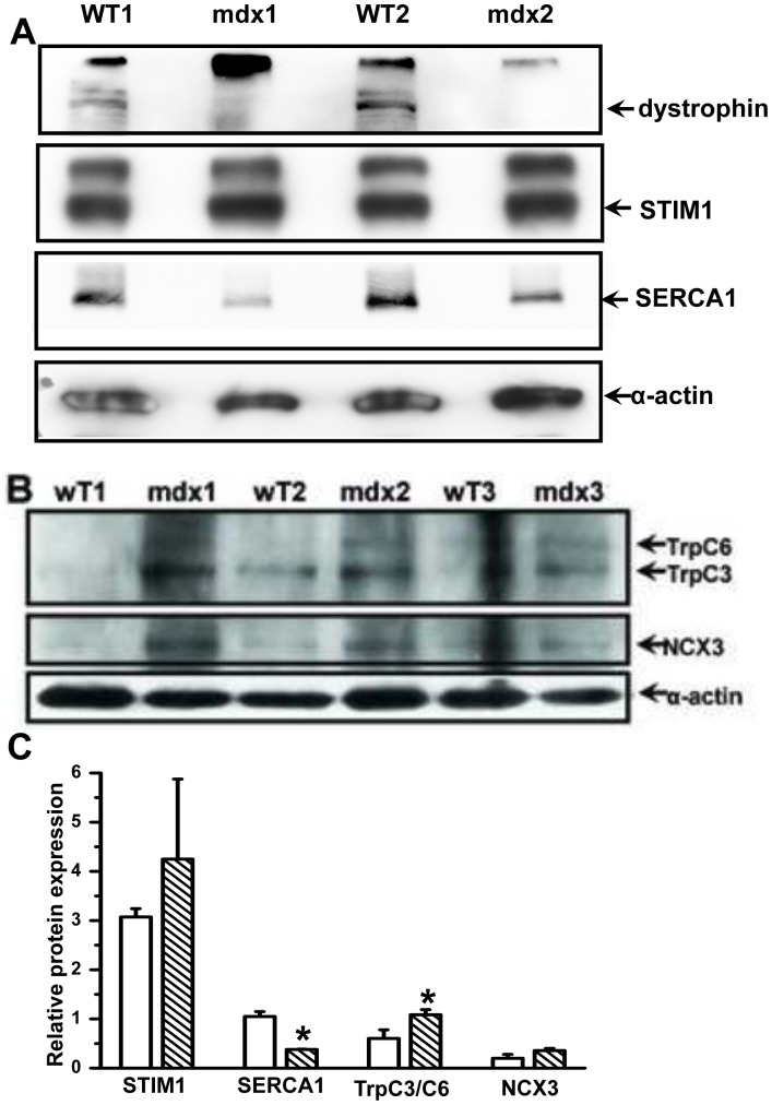
Figure 6. Compensatory change in protein expression of other Ca2+ shuttling pathways in mdx muscles.
(A) Absence of the 427 kDa dystrophin in mdx muscles was confirmed and expression levels of STIM1 and SERCA1 in FDB muscles from the wt C57BL/10ScSnJ and dystrophic C57BL/10ScSn-Dmdmdx/J mice were tested by western blot. Sarcomeric a-actin was used as a loading control. (B) In a separate study, the levels of TRPC3/6 and NCX3 were tested in wt and mdx muscles. (C) Densitometry of detected protein expression relative to α-actin, n = 2 for STIM1 and SERCA1 and n = 3 for TrpC3/C6 and NCX, *P<0.10.

