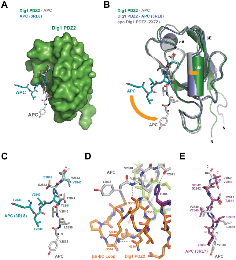
Figure 4. Differential APC peptide binding correlates with PDZ domain conformation change.
A) The Dlg1 PDZ2-APC structure determined in this study with the PDZ2 domain shown in surface representation and colored green, APC peptide shown in stick format and colored grey. Superimposed is the structure of the APC peptide bound to Dlg1 PDZ2 as determined by Zhang et al. (PDB code 3RL8 [26]). The 3RL8 APC peptide is shown in stick format and colored cyan; the PDZ2 domain is not shown for simplicity. B) Ribbon diagram of the Dlg1 PDZ2-APC complex determined in this study (Dlg1 PDZ2 in green, APC in grey), with the structure of the Dlg1 PDZ2-APC complex determined by Zhang et al. (Dlg1 PDZ2 in slate, APC peptide in cyan, PDB code 3RL8) and the apo Dlg1 PDZ2 structure (grey, PDB code 2X7Z [50]) superimposed. The conformation of the Dlg1 PDZ2 domain bound to APC determined by Zhang et al. is more homologous to the Dlg1 PDZ2 apo structure than to the Dlg1 PDZ2-APC structure determined in this study, specifically in the positioning of αB and the βC-αA loop that move outward from the peptide binding cleft upon APC binding. Conformational differences between the PDZ domains and the APC peptides are highlighted by orange arrows. C) Comparison of the APC peptide bound to Dlg1 PDZ2 from Zhang et al. versus this study, oriented as shown in A. The structure of the APC peptide determined in this study use all residues shown to bind the Dlg1 PDZ2 structure. Significant repositioning occurs over residues L2839-T2841 as well as the side chain rotamer positioning of S2842. D) Zoom view of the APC peptide N-terminal region and its interaction with the βB-βC loop. APC and Dlg1 PDZ2 are shown in stick format, colored as in Figure 1C. The backbone region of APC V2840 is stabilized by hydrogen bonds with the N339 side chain. Specific hydrogen bonds are shown as dashed lines. E) Orientation of the APC peptide bound to Dlg1 PDZ2 determined in this study (shown in grey, stick format) versus the APC peptide structure bound to Dlg1 PDZ1, chain J, determined by Zhang et al. (PDB code 3RL7 [26]), colored purple and shown in stick format. APC peptides are shown alone, positioned after structurally aligning the respective PDZ domains they are bound to (not shown).

