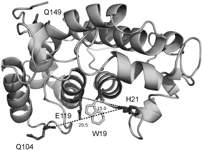
Figure 1. Model structure of NarJT.
The positions of the four residues targeted for cysteine mutations and spin labeling are indicated. The position of the single Trp residue (W19) used for binding affinity measurement is indicated as well as the estimated distances between the Cα of H21 and Q104 or H21 and E119. NarJT is represented in cartoon by Pymol (http://www.pymol.org).

