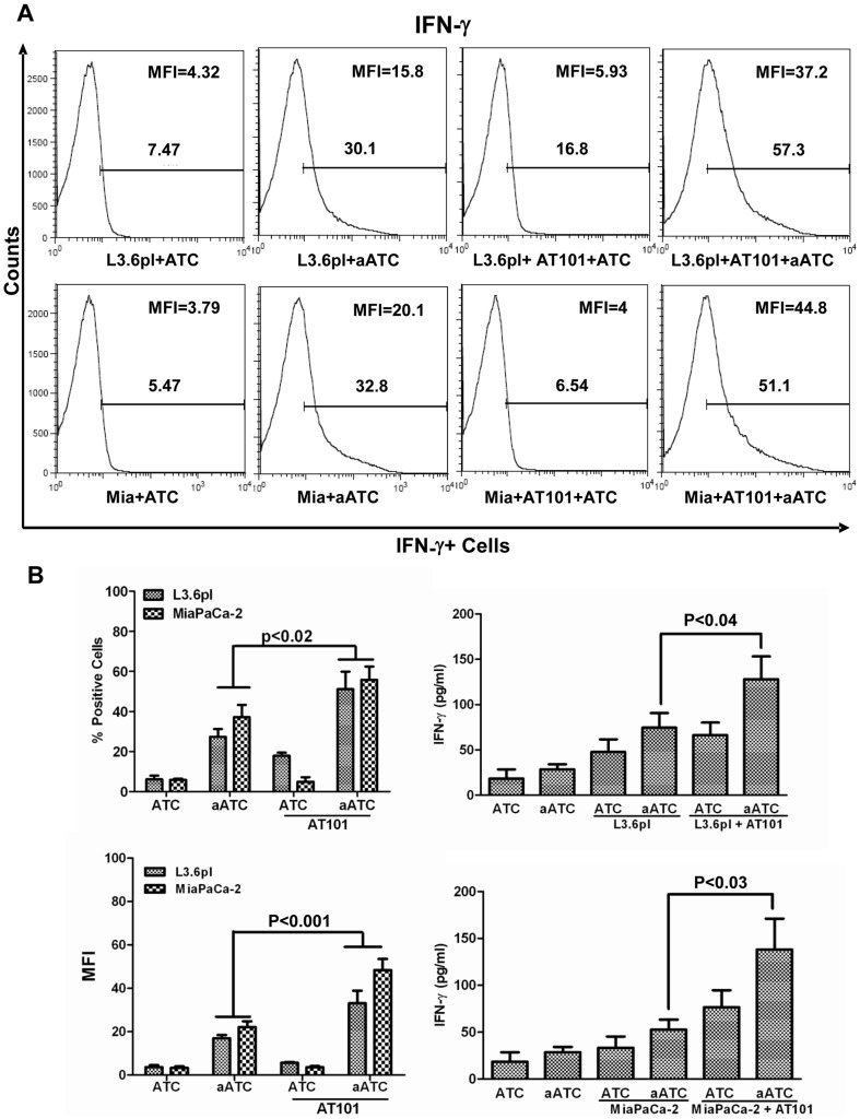
Figure 5. Intracellular staining for IFN-γ by flow cytometry.
MiaPaCa-2 (or Mia) and L3.6pl cells were either pretreated with AT-101 for 24 h or left untreated and a subsequent 4 h incubation with ATC or aATC in the presence of GolgiStop followed by staining for intracellular IFN-γ by flow cytometry. Y-axis shows the counts and the X-axis of each histogram shows the percentage of fluorochrome positive cells. B). Culture supernatants from AT-101 sensitized PC cells co-cultured with ATC or aATC in the absence of GolgiStop were used to detect the secreted IFN-γ in the culture supernatants, and data are presented as mean ± SD from one representative experiment. Y-axis shows the counts and the X-axis of each histogram shows the percentage of fluorochrome positive cells.

