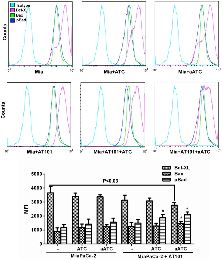
Figure 12. Shows intracellular staining for pBad, Bax and Bcl-XL in MiaPaCa-2 cells were either pretreated with AT-101 for 24 h or left untreated followed by incubation with ATC or aATC before staining.
Top panel show histogram overlays representing isotype control, phosphor-Bad, Bax and Bcl-XL positive cells. Proportion of phosphor-Bad, Bax and Bcl-XL positive cells and MFI were gated on tumor cells. Bottom panel shows the graphic representation of MFI under the indicated experimental conditions.

