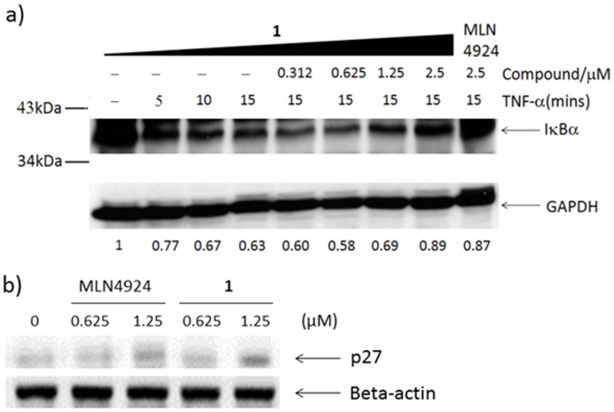
Figure 3. Dose-dependent inhibition of IκBα degradation by 1.
Caco-2 cells were pre-incubated with indicated concentrations of 1 for 16 hours and then stimulated with 5 ng/ml of TNF-α at indicated time intervals. Whole cell lysates were analyzed by Western blot using anti-IκBα antibody. Densitometry estimates of IκBα levels normalized with GAPDH are shown under each lane. b) Caco-2 cells were treated with 1 or MLN4924 for 16 h. The cell lysates were immunoblotted to analyse the level of CRL substrate p27.

