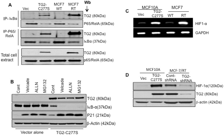
Figure 5. TG2 mediate IκBα degradation via non-proteasomal pathway and results in HIF-1α expression.
A- Total cell extracts prepared from MCF10A-Vec, MCF10A TG2-C277S, drug-sensitive (MCF-7/WT) and drug resistant (MCF-7/RT) MCF-7 cell were immunoprecipitated with anti-p65/RelA or anti-IκBα antibody and immunoblotted with anti-TG2, anti- p65/RelA, or anti- IκBα antibody. Total cell extracts (15 µg protein) were also directly subjected to immunoblotting to determine TG2 and p65/RelA levels. B- MCF10A-Vec and MCF10A TG2-C277S cells were incubated with medium alone (Cont.) or medium containing the indicated proteasomal inhibitor [Bortezomib (20 nM), N-acetyl-leucyl-leucyl-norleucinal (ALLN) (50 µg/mL), and MG-132 (1 µM)] for 2 hours. At the end of incubation, cells extracts were prepared and subjected to immunoblot analysis, using anti-TG2, anti-IκBα and anti-p21 antibody (p21 served as a positive control) and β-actin to ensure equal protein loading. C- RT-PCR analysis showing the basal transcript level of HIF-1α in MCF10A-Vec, MCF10A TG2-C277S, MCF7-WT and MCF7/RT cells. Results shown are from a representative experiment repeated at least 2 times with similar results. D- Immunoblot analysis showing the basal HIF-1α and TG2 protein expression in MCF10A-Vec, MCF10A TG2-C277S, MCF7-WT and MCF7/RT cells under normoxic conditions.

