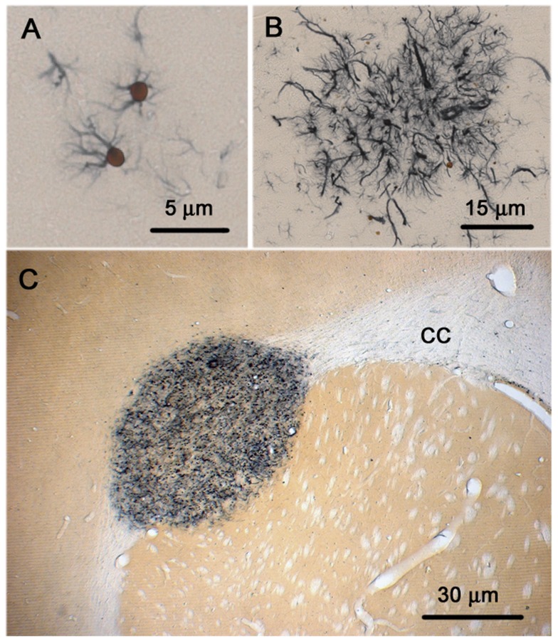
Figure 1. Nestin+ cells in glioma development after in utero ENU exposure.
(A) Characteristic appearance of nestin+ cells that arise in locations commonly noted to harbor tumors at later stages. These usually appear as either a single or more nestin+ cells that arise on a bland background that appears normal on H&E staining. These clusters sometimes can include dozens of cells (B). (C) Small microtumor arising in the external capsule of the corpus callosum (CC). These tumors are hypercellular and apparent on H & E and contain a significant fraction of cells that do not express neural markers.

