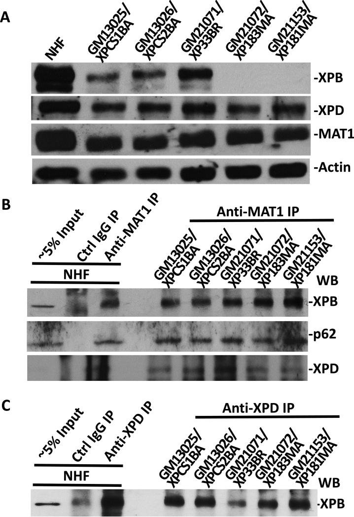
Fig. 2.
Physical association of MAT1 with core TFIIH in XP-B and XP-B/CS fibroblasts. (A) Western blotting of XPB, XPD and MAT1 proteins in NHF, XP-B and XP-B/CS fibroblasts. Protein extracts were made in SDS lysis buffer from indicated normal and XP fibroblasts. The proteins were quantitated and resolved by polyacrylamide gel. The proteins transferred on blots were probed with antibodies to XPB, XPD, MAT1 or β-actin. (B and C) Whole cell extracts were made in RIPA buffer from NHF, XP-B and XP-B/CS fibroblasts. Immunoprecipitation was performed by using control (Ctrl) or MAT1 (B) or XPD (C) antibodies. The immunoprecipitates were analyzed by Western blotting with specific XPB, p62 or XPD antibodies.

