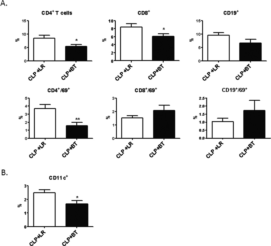
Figure 9. Percentage of circulating leukocytes in septic animals after resuscitation with PRBCs or Ringer’s lactate.
Blood was collected from septic mice one day after receiving either allogeneic PRBC (n=11) or LR (n=9) in heparinized syringe by intra-cardiac puncture. Leukocytes were analyzed by flow cytometry using anti-CD4 (T-helper cells), anti-CD8 (cytotoxic T lymphocytes), and anti-CD19 (B cells). The activation status was determined by using anti-CD69 (A). Dendritic cells were identified using anti-CD11c (B). Data shown is the mean from three separate experiments (*p<0.05; **p<0.01).

