
Figure 7. Pre-stimulated CD8+ T cells from patients on HAART eliminate CD4+ T cells infected with autologous viruses.
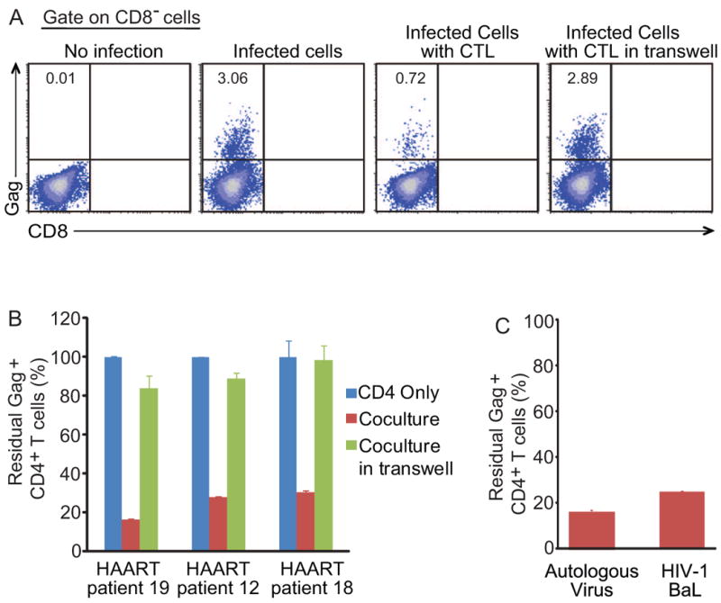
(A and B) PBMCs from patients on HAART were isolated and cultured in basal medium for 5-6 days to allow the decay of remaining intracellular triphosphorylated nucleoside/nucleotide reverse transcriptase inhibitors, and then stimulated with phytohemagglutinin. CD4+ T cells were isolated 2 days after stimulation and infected with autologous viruses. Pre-stimulated autologous CD8+ T cells were added into the culture 4 hours after infection at the effector-to-target ratio 1:1. Raltegravir and enfuvirtide were added in the culture to prevent further rounds of viral replication. The fraction of residual Gag+ CD4+ T cells was measured after 3 days of co-culture. Numbers in the quadrant indicate the percentage of cells. Data represent the mean ± SEM, N=3. (C) Patient autologous viruses or HIV-1 BaL were used to infected CD4+ T cells. Infected cells were then cocultured with CD8+ T cells as described above. The fraction of residual Gag+ CD4+ T cells was measured after 3 days of co-culture. Data represent the mean ± SEM, N=3.