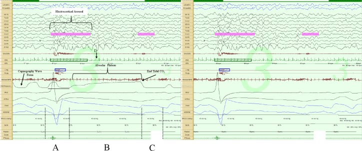
Figure 1. Prolonged expiratory apnea, 30-sec epoch.
The image on the left includes the capnography tracing, while the one on the right does not. Augmented breath (A); respiratory pause (B); first post-pause breath (C) in the direction of exhalation (positive polarity); lowest instantaneous heart rate during the respiratory pause (D). Note the prolonged alveolar plateau phase in the capnopgraphy tracing. Note there is about a 3- to 5-sec sampling delay in the capnography tracing, as illustrated by the horizontal line below the tracing.

