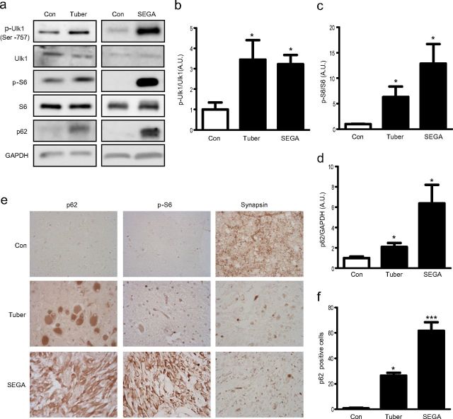
Figure 4.
Autophagy is inhibited in the brains of TSC patients. a, Representative Western blots from control cortex from normal human brains compared with cortical tuber or SEGA tissues from human TSC patients. Quantifications of phospho-Ulk1 (b), phospho-S6 (c), and p62 (d) in control, cortical tuber, and SEGA. e, Immunohistochemical staining for p62, phospho-S6, and synapsin in human control cortex and tissues from TSC patients, including tuber and SEGA (40× magnification). f, Quantifications of p62-positive cell counts from 20× magnification (mean ± SEM; n = 3–6; t test or ANOVA with Tukey's post hoc test; *p < 0.05; **p < 0.01; ***p < 0.001). Con, Control; Tuber, cortical tuber.

