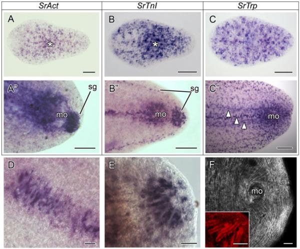
Figure 2.
SrAct, SrTnI, and SrTrp gene expression patterns in the acoel Symsagittifera roscoffensis. Anterior is toward the left in all aspects. (A) SrAct expression in a juvenile. The positive cells appear scattered along the AP axis of the juvenile. The highest concentration of actin-expressing cells is around the mouth region (asterisk), where the mouth accessory muscles and U-shaped muscles are present. (A”) SrAct expression in the posterior tip of an adult. The expression is restricted to the male genital opening (mo), the region frontal to it, and the muscle mantles of sagittocysts (sg). (B) SrTnI expression in a juvenile specimen. The expression of the troponin I gene appears scattered with the highest concentration of positive cells in the mouth region (asterisk). (B”) SrTnI expression in the posterior part of an adult. The expression of SrTnI is recovered in the region of the male gonopore (mo), in a longitudinal band anterior to it, and in the muscle mantles of sagittocyts at the posterior-most tip and in two lateral longitudinal bands (sg). (C) SrTrp expression in a juvenile. Contrary to the SrAct and the SrTnI genes, SrTrp is more broadly expressed along the body of the juvenile. The frontal region has a greater number of SrTrp-positive cells if compared with SrAct and SrTnI. (C”) SrTrp expression in the posterior part of an adult. As in the juvenile, the SrTrp-positive cells are more scattered than SrAct and SrTnI ones. High expression levels are seen in the region of the male gonopore and in longitudinal bands anterior to it (arrowheads). (D) Detail of the muscle mantles of the sagittocysts in the region between the female and the male genital opening as revealed by the anti-SrTnI riboprobe. (E) Detail of the muscle mantles of the saggitocysts, revealed by the anti-SrTnI riboprobe, at the posterior-most tip of an adult specimen. (F) Anti-SrTrp antibody staining in the caudal end of an adult. The antibody detects all types of muscles, except for the muscle mantles that surround the sagittocysts. The latter, labeled by the phalloidin, are shown in the inset. Scale bars: A-C 50 μm, A”–C” 100 μm, D–F and inset 25 μm. mo, male genital opening; Sg, muscle mantle of the sagittocyst.

