
Figure 1.
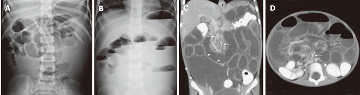
Radiologic study. A, B: Plain abdominal radiography in supine (A) and upright (B) positions showed small-bowel dilatation with multiple air-fluid levels in the upright position (B); C: Coronal view of the abdominal computed tomography (CT) scan revealed twist of the mesentery and its vessels (arrows) with a fluid-filled multiloculated mass in the right lower quadrant (between arrowheads); D: Axial view of the abdominal CT scan showed a twist of the mesenteric fat and its vessels (arrow) with small-bowel dilatation.