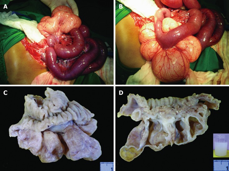
Figure 2.

Gross pathology. A, B: Intraoperative photographs show a lobulated cystic yellowish pink mass that originated from the small-bowel mesentery (arrow in A) with small bowel dilatation. The mass shows vascular streaks (B); C, D: The formalin-fixed specimen reveals a partially collapsed, cystic, lobulated, pale tan mesenteric mass with vascular streaks (C). The cut surface reveals thin-wall multicystic spaces with thickened white appearance in some cystic walls (D). The inset shows milky white fluid. The small-bowel mucosa is not remarkable.
