
Figure 4.
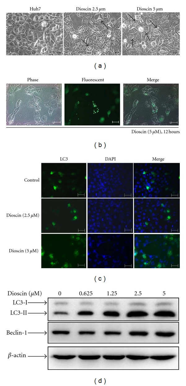
Autophagy was induced in dioscin-treated cells. Huh7 cells were treated with 2.5 and 5 μM dioscin for 12 hours and a microscopic observation revealed the formation vacuoles in cytoplasms of treated cells (a). After a successful transfection with pEGFPC1-LC3, Huh7 cells were treated with dioscin of 5 μM for 12 hours, followed by an observation for LC-3 (green fluorescence) under fluorescence microscopy (b). After a 24-hour treatment with dioscin, DAPI staining and subsequent fluorescence microscopy for LC-3 and DAPI (blue fluorescence) were conducted (c), as well as western blotting with antibodies against LC3 and beclin-1 (d). Scale bars = 100 μM.