Figure 1.
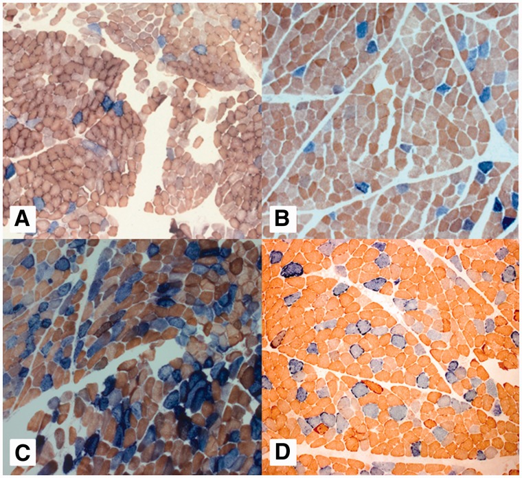
Mitochondrial histochemical changes associated with RRM2B mutations. Representative sequential COX-SDH histochemistry demonstrates a mosaic distribution of COX-deficient muscle fibres (blue) among fibres exhibiting normal COX activity (brown). Illustrated are the images for (A) Patient 5, (B) Patient 10, (C) Patient 19 and (D) Patient 20. Patients 5 and 10 have autosomal-dominant RRM2B mutations and a milder histochemical COX defect compared with Patients 19 and 20 (autosomal-recessive RRM2B mutations), in whom a more severe biochemical defect is clearly apparent.

