Figure 2.
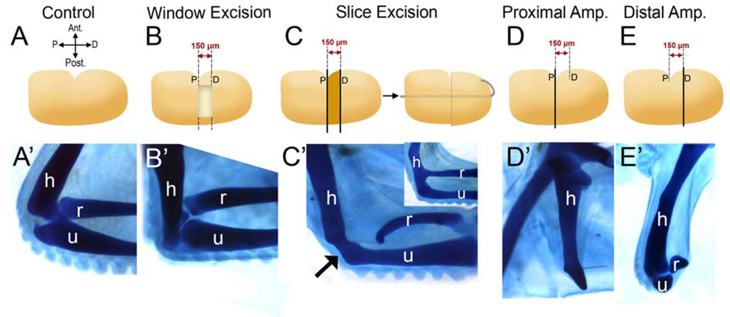
The elbow joint of the chicken embryo is able to regenerate depending on the method of joint removal. A) The contralateral limb served as a control. (A’–E’) All samples were stained with Victoria blue to visualize the cartilage morphology. B) Window Excision (WE). Using the anterior indentation as a landmark, a 150µm-wide block of tissue containing the prospective elbow is removed leaving a “window”. C) Slice Excision (SE). Using the anterior indentation as a landmark, a piece of the limb, 150µm-wide containing the prospective elbow is sliced out and discarded. The remaining distal piece of the forelimb is pinned onto the stump, leaving no space between the two wound surfaces. Both surgeries were performed at st. 26 and embryos were incubated for four days following surgery. B) After WE surgery, 10 out of 20 limbs displayed joint regeneration (B’). When the joint was removed by SE surgery (C), regeneration did not take place (n=6) and a fused cartilage was observed (C’, arrow). Fusion is observed mainly between the humerus (h) and the ulna (u); however, h-u and h-r (r: radius) fusion together is also observed (C’, inset). D and E) Proximal and distal amputations verify that the elbow was removed by both WE or SE methods. Limbs were amputated at the indentation, indicated by “P” for proximal (D) or 150 µm distal to the indentation (E) indicated by “D” for distal amputation (see lines in D, E). The proximal amputations did not include elbow tissue and only the humerus could be observed (D’). On the other hand, the distal amputations contained the elbow tissue (E’). h: humerus, r: radius, u: ulna.

