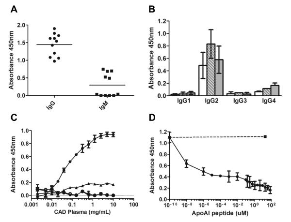
Figure 3. Screening for circulating immunoglobulins that recognize 3-nitrotyrosine.
Panel A: IgG and IgM levels in human plasma were measured by ELISA in ten randomly selected CAD plasma subjects, using the 3-nitrotyrosine octapeptide to bind the immunoglobulins and the respective anti-human immunoglobulin for detection. Panel B: IgG subclass was determined by ELISA in three independent CAD plasma samples, using nitrated ceruloplasmin (white bars), nitrated fibronectin (dashed bars) and nitrated fibrinogen (grey bars) as capturing antigen, and HRP-conjugated mouse anti-human IgG 1, IgG2, IgG 3, and IgG 4 for detection. Panel C: Typical antigen-antibody binding curves from 3 randomly selected CAD subjects each performed in triplicate, using as ligand the 3-nitrotyrosine-octapeptide conjugated to HRP (●). No binding was observed using the same tyrosine-containing octapeptide conjugated to HRP (○). The binding of the 3-nitrotyrosine-octapeptide conjugated to HRP was eliminated with 75 μM unlabeled 3-nitrotyrosine-octapeptide (▴) or by 250 μM 3-nitrotyrosine (∎). Panel D: The binding of the 3-nitrotyrosine-octapeptide conjugated to HRP to 3 different plasma samples, each performed in triplicate, was also eliminated with increasing concentrations of apolipoprotein A-I peptide 213-219 (213L-A-E-nitroY-H-A-K219) which contained 3-nitrotyrosine in position 216 (●) but not by the same peptide that contained tyrosine in position 216 (∎). Data reports mean ± standard deviation.

