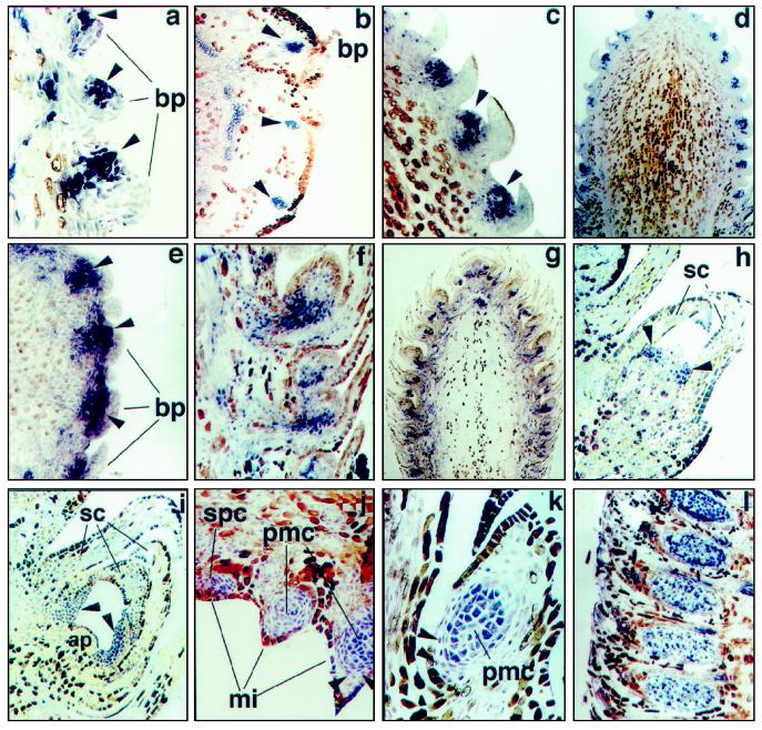
Figure 4.
In situ localization of PrMADS3 transcripts in developing SCBs, PCBs, and DSBs. a, Differentiating SCB. Expression in the group of cells initiating ovuliferous scale primordia is shown by arrowheads (magnification ×42). b, Cross-section of differentiating SCB. Expression in the group of cells initiating ovuliferous scale primordia is shown by arrowheads (magnification ×45). c and d, SCB at stage 2. Ovuliferous scale primordia shown by arrowheads (magnification ×100 and ×11.25, respectively). e, Cross-section of SCB at stage 3. Ovuliferous scale primordia shown by arrowheads (magnification ×55). f and g, SCB at stage 3 (magnification ×45 and ×8.3, respectively). h, DSB with initiating needle primordia (arrowheads) (magnification ×90). i, DSB with developing needle primordia (arrowheads) (magnification ×45). j and k, Differentiated PCB with initiating microsporophylls (stage 1). Two layers of tapetal cells surrounding pollen mother cells are shown by arrowheads (magnification ×45 and ×60, respectively). l, PCB after completion of microsporophyll initiation (stage 2) (magnification ×23.5). All sections except b and e, which are cross-sections, are longitudinal. ap, Apical meristem; bp, bract primordia; mi, microsporophylls; pmc, pollen mother cells; sc, sterile cataphylls; and spc, sporogenous cells.

